


Cavernoma and colloid cyst; They are two different structural disorders located in the brain tissue, which although they are usually benign, can have serious neurological consequences due to their critical location.
Cavernomas are caused by a vascular anomaly, while colloid cysts are cystic formations that develop in the circulatory tract of cerebrospinal fluid.
What is Cavernoma (Cavernous Malformation)?
Cavernoma is a vascular ball in the central nervous system, formed by thin-walled and enlarged vessels, resembling a “berry” appearance.
Since these structures lack normal vascular tissue, blood flow through them is very slow and their walls are quite fragile.
These formations, also called “cavernous angioma” or “cavernoma” in the medical literature, are not tumors, but a structural vascular malformation.
Although they are mostly located in the cerebral hemispheres, they require high surgical precision when they are located at vital points such as the brain stem or spinal cord.
What are the Symptoms of Cavernoma?
Cavernomas can sometimes remain silent for years without any symptoms and can be detected incidentally in MRI (Magnetic Resonance) images taken for another reason.
However, when they leak into the surrounding brain tissue at a microscopic level or perform a larger hemorrhage, the following symptoms occur:
- Epileptic Seizures: It is the most common symptom of cavernoma. Iron accumulation (hemosiderin) in the surrounding brain tissue stimulates nerve cells, triggering seizures.
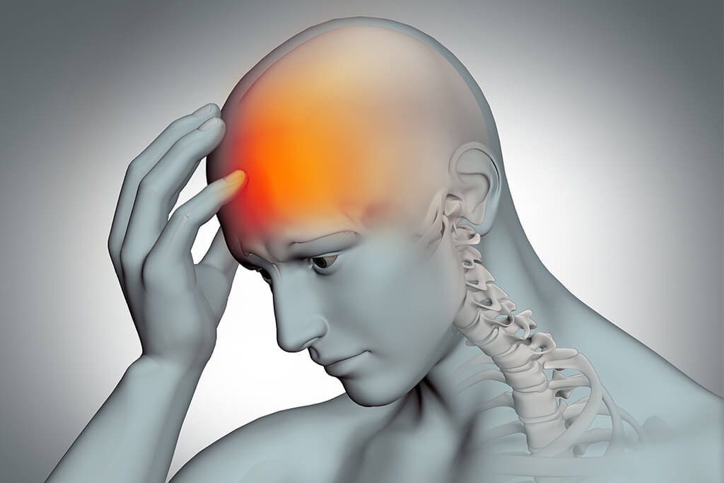
- Severe and Persistent Headaches: Pain that develops due to bleeding or mass effect and is difficult to relieve with medication.
- Neurological Losses: Varies according to the location of the tumor; weakness, numbness, visual disturbances, or difficulty speaking in the arms or legs.
- Balance Disorder and Double Vision: These are symptoms that seriously affect the quality of life, especially seen in cavernomas located in the brain stem.
- Sudden Worsening Due to Bleeding Risk: The bleeding risk of a cavernoma is around 1%-2% annually; However, cavernoma that bleeds once has an increased risk of bleeding again.
Assoc. Prof. Dr. Erdinç Özek; “The most critical point in cavernoma follow-up is ‘active follow-up’. If a cavernoma is detected by chance and is silent, it can only be traced. However, if the patient has started a seizure or has caused neurological loss by bleeding, that mass has now become a surgical target. When making a decision, it is necessary to put the natural history of the cavernoma and surgical risks on the scales.”
Cavernoma Diagnostic Methods
Cavernomas cannot be clearly diagnosed with a standard brain x-ray or often Computed Tomography (CT).
The “gold standard” in diagnosis is Magnetic Resonance Imaging (MRI).
- Magnetic Resonance (MRI): The characteristic “popcorn” or “berry” appearance of the cavernoma is most clearly detected by MRI.
- Gradient Echo (GRE) and SWI Sequences: These special MRI techniques detect old bleeding traces (hemosiderin) around the cavernoma, allowing small and hidden cavernomas to be seen as circular black rings.
- Angiography (DSA): Cavernomas are “angiographically occult” lesions. In other words, they are not seen in angiography taken by giving vascular dye. This feature is the most important difference that distinguishes them from other vascular tangles (AVMs).
Cavernoma Treatment Methods
Treatment plan; It is individualized based on the size of the cavernoma, the bleeding history, the age of the patient, and how deep in the brain the lesion is.
Observation and Follow-up
If the cavernoma is detected incidentally, does not cause any complaints and does not put pressure on a vital area, the “wait-and-see” strategy is applied.
MRI images taken at regular intervals check the size of the cavernoma and whether there is a new leak around it.
Microsurgical Method
The definitive and permanent treatment for cavernoma is complete surgical removal.
- Indication: It is applied in cavernomas that have bleeding more than once, cause drug-resistant seizures or cause neurological losses.
- Technique: Under high-magnification surgical microscopes, the mass is removed without damaging the surrounding brain tissue.
- Neuronavigation: Especially in deep-seated lesions, navigation technology similar to “GPS” is used to enter from the safest point of the brain.
Gamma Knife Radiosurgery
It is an alternative for cavernomas in areas that are too risky to reach surgically (e.g. deep brainstem locations).
- Purpose: It aims to reduce the risk of bleeding by disrupting the vascular structure of the cavernoma with targeted intense radiation therapy.
- Note: Radiosurgery does not completely destroy the cavernoma; It shows its effect in a long period of 2-3 years and may not offer as definitive a solution as surgery.
According to Assoc. Prof. Dr. Erdinç Özek; “Our basic rule in cavernoma surgery; It is also to carefully evaluate the ‘hemosiderin ring’ around the lesion. Especially in patients with seizures, it may not be enough to remove the cavernoma; Proper cleaning of this dyed tissue (iron deposit) increases seizure control success to 80-90%. However, protecting healthy brain tissue during this procedure is our top priority.”
| Feature | Observation and Follow-up | Microsurgery | Gamma Knife |
| Hospital Stay | Not required | 3 – 5 Days | Same Day |
| Final Solution | No (Follow-up-oriented) | Yes (If Full is subtracted) | Partial (Reduces risk) |
| Risk Factor | Bleeding monitoring is required | Surgical risks | Radiation effect |
| Application Area | Silent lesions | Bleeding/Seizure | Unreachable deep areas |
What is a Colloid Cyst?
Colloid cyst is a type of cyst that is located in the midline of the fluid-filled cavities called ventricles in the brain (especially at the ceiling of the third ventricle), benign but life-threatening in terms of location.
This cyst is filled with a jelly-like dark fluid (colloid) and is surrounded by a thin capsule.
This structure, which accounts for about 1% of all brain tumors, is not actually a cancer; however, because it sits right above the brain’s water flow pathways (Monro holes), it can act as a “plug” and abruptly stop the flow of cerebrospinal fluid (CSF).
What are the Symptoms of Colloid Cyst?
Symptoms of colloid cysts are usually associated with increased intracranial pressure (hydrocephalus) due to obstruction of the flow of cerebrospinal fluid.
- Sudden and Severe Headache: Pain that usually comes in attacks, can change with the position of the head, and intensifies when leaning forward.
- Nausea and Gushing Vomiting: It is the clearest indicator of increased intracranial pressure.
- Cognitive Issues: Memory loss, distraction and confusion.
- Visual Disturbances: Double vision or blurred vision.
- Sudden Fainting or Falling Episodes: Sudden emptying of the legs and falling to the ground without loss of consciousness (“drop attacks”).
- Risk of Sudden Death: Although rare, sudden deaths due to brain herniation as a result of sudden and complete obstruction of the waterway by the cyst have been reported in the literature.
Assoc. Prof. Dr. Erdinç Özek; “The most insidious feature of colloid cysts is that the symptoms are positional. When the patient holds his head at an angle, the cyst blocks the waterway and severe pain begins; When the head is moved, the cyst may move and open the waterway and the pain goes away suddenly. ‘Temporary’ headaches should be taken seriously as this can lead to delays in diagnosis.”
Colloid Cyst Diagnosis and Diagnosis
The most sensitive methods in the diagnosis of colloid cysts are radiological imaging techniques. The density and size of the cyst directly affect the treatment decision.
- Computed Tomography (CT): Colloid cysts usually appear brighter (hyperdense) than the surrounding tissue on CT. This is a sign that the fluid inside the cyst is too dense.
- Magnetic Resonance (MRI): It is the most important pillar of diagnosis. MRI shows with millimeter precision how much the cyst narrows the cerebrospinal fluid pathways and whether there is a collection of water in the brain (hydrocephalus).
- MR Spectroscopy: It is used when necessary to understand the content of the cyst and its difference from other cystic masses.
Clinical Experience Note (Anonymous Case): A 35-year-old male patient presented with stabbing headaches that increased when leaning forward for the last 2 weeks and lasted for a few minutes. In the brain MRI examination, a colloid cyst with a diameter of 12 mm was detected at the entrance of the third ventricle, narrowing the waterways. Upon observing mild hydrocephalus (water accumulation in the brain) in the patient, an emergency intervention decision was made and treatment was planned with the closed method.
Colloid Cyst Treatment Methods
The treatment decision for colloid cysts is made according to the size of the cyst, the degree to which it obstructs the flow of cerebrospinal fluid, and the patient’s symptoms.
If the cyst is small and does not obstruct the waterways, only close follow-up may be recommended; However, in cases where there is a risk of blockage, surgical intervention is vital.
Microsurgical Excision
It is a traditional and proven method. The cyst is reached using the natural cavities of the brain or by opening a microscopic corridor.
- Application: Under high-magnification surgical microscopes, the capsule of the cyst is separated from the surrounding tissues and completely removed along with its contents.
- Advantage: It has the highest success rate in removing the cyst completely (total excision); This reduces the risk of recurrence of the cyst to almost zero.
- Disadvantage: It may require a slightly larger surgical incision than the endoscopic method.
Endoscopic Colloid Cyst Surgery
It is a minimally invasive (closed) surgical method that is considered the “gold standard” today.
- Technique: It is entered through a small hole of about 1-2 centimeters in the skull with an endoscope with a high-resolution camera at the end.
- Process: The surgeon drains the contents of the cyst with the image watched on the monitor and then removes the cyst wall.
- Advantage: Since minimal intervention is made to the brain tissue, the healing process is very fast, the risk of bleeding is low and the suture scar is minimal in terms of aesthetics.
Postoperative Recovery Process
Thanks to modern surgical techniques, patients can return to their normal lives in a short time, both physically and neurologically.
- First 24 Hours: The patient is usually observed in the intensive care unit or close follow-up unit. Neurological functions are checked hourly.
- Mobilization: The day after surgery, patients are usually stood up and walked.
- Hospital Stay: Patients can usually be discharged within 2-3 days for endoscopic surgery and within 4-5 days for microsurgery.
- Return to Normal Life: It usually takes between 2 and 4 weeks for the stitches to heal and for the patient to return to work at full capacity.
- Tracking: With control MRIs taken in the 3rd or 6th month after surgery, it is confirmed that the cyst is completely cleared and the intracranial pressure has returned to normal.
Frequently Asked Questions
What happens if a colloid cyst is not operated on?
If the cyst suddenly blocks the waterway, cerebrospinal fluid accumulates and intracranial pressure rises suddenly. This can lead to coma or, unfortunately, sudden death. For this reason, risky cysts are considered as “time bombs”.
Does the cyst recur after surgery?
If the cyst wall (capsule) is completely removed by microsurgery or a successful endoscopic intervention, the risk of recurrence is extremely low.
Can cavernoma and colloid cyst go away with medication?
Both conditions are structural disorders (vascular ball and cystic sac). Medications are only used to control symptoms such as headaches or seizures, they cannot destroy the mass itself.
Resource and Expert Knowledge
This content has been prepared with Assoc. Prof. Dr. Erdinç Özek’s current clinical data and extensive case experience in 2026 on neuroendoscopy, minimally invasive neurosurgery, and vascular malformations. Assoc. Prof. Dr. Erdinç Özek is a neurosurgeon specializing in the treatment of complex brain masses with advanced technological equipment.