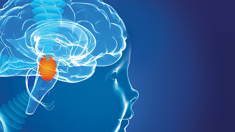


Brain and spinal cord tumors are masses that form as a result of uncontrolled and abnormal proliferation of cells in the tissues that make up the central nervous system.
These tumors can cause complex neurological pictures by pressing on or infiltrating the nerve tissues that control vital functions, depending on the area where they are located.
What are Brain and Spinal Cord Tumors?
Central nervous system tumors include masses that develop by spreading from brain tissue (primary) or from another part of the body (metastatic).
Since the brain and spinal cord are closed spaces surrounded by bone structures (skull and spine), any growing mass, even if they are benign, can cause serious damage by increasing intracranial pressure or compressing the spinal cord.
What are the Symptoms of Brain and Spinal Cord Tumor?
Symptoms of central nervous system tumors are usually directly related to the function of the region where the tumor is located.
In general, symptoms that occur as a result of increased intracranial pressure or narrowing of the spinal canal can have an insidious onset or manifest themselves with sudden seizures.
Brain Tumor Symptoms
Symptoms of brain tumors develop due to the edema and space-occupying effect of the mass on the brain tissue.
- Persistent and Severe Headache: Pain that is more pronounced especially in the morning and increases in frequency and intensity over time.
- Nausea and Vomiting: Nausea, usually in the morning, gushing and accompanied by pain.
- Seizures (Epilepsy): The first episode of fainting or convulsions in an adult with no previous history of seizures.
- Personality and Behavioral Changes: Memory problems, irritability or sudden emotional fluctuations seen in frontal lobe tumors.
- Vision and Speech Disorders: Double vision, narrowing of the visual field, or difficulty choosing words.
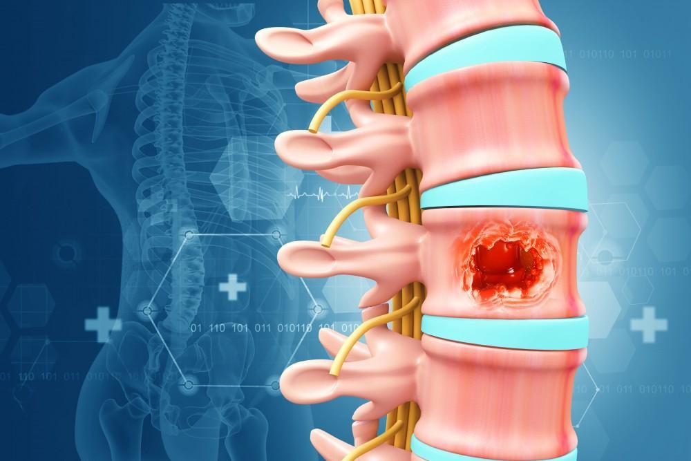
Spinal Cord Tumor Symptoms
Spinal cord tumors usually give symptoms of “interruption” that affect the lower parts of the body because they cut or compress the nerve conduction line.
- Back and Low Back Pain: Pain that intensifies at the level of the tumor, increases at night and does not go away with rest.
- Sensory Loss: Burning, numbness, tingling and reduced sensation of hot and cold in the hands or feet.
- Motor Weakness: Loss of strength in the legs or arms, shortening the walking distance or making it difficult to carry items.
- Loss of Balance and Coordination: A feeling of wobble left and right or tangled legs while walking.
- Reset Reflexes: Severe loss of function such as loss of urinary or fecal control in advanced stages.
Clinical Experience Note (Anonymous Case): In a 45-year-old patient who presented with only severe low back pain and was followed up with suspicion of hernia, it was determined that the cause of the pain was a benign but space-occupying tumor originating from the spinal cord membrane (meningioma) as a result of the contrast MRI examination. After complete removal of the tumor by microsurgery, all neurological complaints of the patient regressed within 48 hours.
Causes and Risk Factors of Brain and Spinal Cord Tumors
Although the exact cause of central nervous system tumors cannot be fully determined in most cases, genetic mutations at the cellular level are known to trigger this process.
Unlike the competitors, it should be noted that; Not only external factors, but also disruptions in the cells’ own repair mechanisms play a key role in tumor formation.
- Genetic Factors: Inherited conditions such as neurofibromatosis (Types 1 and 2), Von Hippel-Lindau disease, and Li-Fraumeni syndrome increase the risk.
- Radiation Exposure: Ionizing radiation (radiotherapy) applied to the head or spine area for another reason in the past can trigger the development of a secondary tumor years later.
- Immune System Problems: Some brain lymphomas are more common in immunocompromised people (HIV or after organ transplantation).
- Environmental Factors: Although some industrial chemicals have been investigated, no proven direct link has been established between cell phone or base station use and tumor development today.
Classification of Brain and Spinal Cord Tumors
The World Health Organization (WHO) classifies these tumors from Stage 1 to Stage 4 based on the degree of aggressiveness of the cells.
Benign Tumors
Benign tumors grow slowly and are usually separated from the surrounding brain tissue by clear boundaries.
However, the term “benign” does not mean that these tumors are harmless. Since the skull and spinal canal are a limited space, these masses can press on vital centers as they grow, leaving permanent damage. After complete surgical removal, the probability of recurrence is generally low.
Malignant Tumors
Malignant tumors grow rapidly and spread by taking root (infiltration) in the surrounding tissues.
The boundaries of these masses are unclear; Separation of healthy tissue and tumor tissue during surgery requires high precision. Treatment processes usually include oncological protocols in addition to surgery.
According to Assoc. Prof. Dr. Erdinç Özek; “Our patients usually focus on the question ‘is my tumor benign or malignant?’ However, what is really important for us is the ‘location’ of the tumor, that is, where it is. A 1 cm benign tumor in a vital center may be more surgically challenging than a large mass in a quiet area.”
Diagnostic Methods
Modern diagnostic methods no longer clarify only the presence of the tumor, but also the biochemical structure of the cells and which areas (speech, walking centers) they come into contact with before the surgeon.
- Contrast MRI (Magnetic Resonance): It is the cornerstone of diagnosis. Thanks to the drug given intravenously, the boundaries of the tumor and the level of blood supply are clearly seen.
- Functional MRI (fMR): It allows us to draw the “safe surgical boundary” by determining the location of speech or movement centers before surgery.
- MR Spectroscopy: By analyzing the biochemical content of the tumor, it provides information about the stage of the tumor without performing a biopsy.
- PET/CT: It measures the metabolic activity of the tumor; It is used to find the main source, especially in metastatic tumors.
- Biopsy: It is the process of removing a small piece for a definitive diagnosis. It is performed with neuronavigation technology with a millimeter margin of error.
Clinical Experience Note (Anonymous Case): In a patient who came with headache and mild forgetfulness, although the MRI image indicated a malignant tumor (Glial tumor), MR Spectroscopy and subsequent stereotactic biopsy revealed that the condition was actually a rare inflammatory reaction; The patient was saved from unnecessary open surgery and recovered with medication.
Brain and Spinal Cord Tumors Treatment Methods
In modern medicine, the treatment of brain and spinal cord tumors is planned with a “multidisciplinary” approach.
When making a treatment decision, the location, type, stage of the tumor and the general health status of the patient are evaluated as a whole.
Goal; It is not only to ensure tumor control, but also to keep the patient’s quality of life and neurological integrity at the highest level.
Surgical Treatment (Resection)
Surgery is the first and most critical step of treatment for most brain and spinal cord tumors.
The principle of “maximum safe resection” aims to remove the tumor at the highest possible rate without damaging healthy tissues.
- Neuronavigation: During the surgery, it guides the surgeon like a GPS and determines the boundaries of the tumor with millimeter precision.
- Neuromonitoring: By monitoring nerve conduction throughout the surgery, it minimizes the risk of damage to critical areas (speech, walking).
- Ultrasonic Aspirator: It cleans the tumor tissue by breaking it down and absorbing it without damaging the surrounding tissues.
Radiotherapy (Radiation Therapy)
High-energy beams are used to destroy any remaining microscopic tumor cells after surgery or to control tumors that are not suitable for surgery.
Thanks to advanced technologies (CyberKnife, Gamma Knife), the rays are focused only on the tumor tissue and the surrounding brain tissue is protected.
Chemotherapy (Drug Therapy)
They are drugs used to stop cell division in malignant tumors.
Special agents that can cross the brain barrier are preferred to slow down the growth rate of the tumor.
Smart Medicine and Immunotherapy Options
In the medical world of 2026, “targeted therapies” designed according to the genetic profile of the tumor are at the forefront.
Immunotherapy, on the other hand, offers promising results in cases where traditional methods are insufficient by training the patient’s own immune system to recognize and destroy tumor cells.
| Treatment Method | Main Purpose | Application Process |
| Surgery | Physical removal of the mass | 3-5 days hospitalization |
| Radiotherapy | Destruction of the remaining cells by radiation | In weekly sessions |
| Chemotherapy | Systemic control and cell arrest | Either orally or intravenously |
| Smart Medicines | Attack on genetic targets | Tumor genetic planning |
According to Assoc. Prof. Dr. Erdinç Özek; “The success of the surgery cannot be measured only by removing the tumor. The technologies we use during surgery, such as neuromonitoring, are to ensure that the patient returns to his old life, loved ones and work at the same neurological level after surgery.”
Recovery Process After Brain and Spinal Cord Tumor Surgery
The healing process varies from person to person depending on the extent of the surgery and the location of the tumor.
- Initial Observation: Patients are usually monitored in the intensive care unit for one night after surgery.
- Mobilization: The patient is allowed to walk and feed the day after the surgery.
- Rehabilitation: If there is a loss of strength due to the tumor, starting physical therapy in the early period accelerates recovery.
- Controls: According to the pathology result, additional treatments (radiation or medication) are planned and MRI follow-ups are performed at regular intervals.
Frequently Asked Questions
Does personality change after brain surgery?
It depends on the location of the tumor and the precision of the surgery. It is our priority to perform the procedure with modern microsurgical methods without damaging the emotional and personality centers.
Should every tumor be operated on?
Some benign tumors that are very small, found incidentally and do not grow can only be followed up with the “wait and watch” method.
Is it possible to stay awake during surgery?
Yes. With the “Awake Craniotomy” technique, surgery is performed by talking to the patient in tumors very close to the speech center; Thus, the ability to speak is fully preserved.
Resource and Expert Knowledge
This article has been prepared in light of Assoc. Prof. Dr. Erdinç Özek’s neurooncological surgery protocols and current treatment algorithms for 2026. Assoc. Prof. Dr. Erdinç Özek is a senior surgeon with deep experience in microsurgical techniques and neuronavigation-assisted interventions in complex brain and spinal cord tumors.